 |
Using the Microscope. Basic Tutorial. Setting Up Köhler Illumination. |
Part 4 of 9 Page 1 of 1 |
|
Using the Microscope. Basic Tutorial. Setting Up Köhler Illumination. |
Part 4 of 9 Page 1 of 1 |
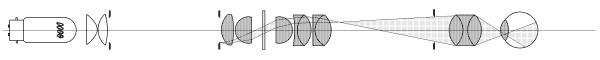
The following account assumes a laboratory grade microscope with a mirror and a separate free-standing microscope lamp with variable power supply. Users of microscopes with built-in illumination must adapt the instructions where necessary and be content with the manufacturer's setting when no means of adjustment is available. Whilst an adjustable mirror can be the cause of maladjustment, it also provides the experienced microscopist with a valuable means of making fine adjustments to the system, and is a useful tool in troubleshooting illumination problems. A separate microscope lamp and mirror also allows the easy placement of light-modifying filters and diffusers etc. in the illumination path. The instructions can be implemented without problems for dry objectives in the x10 - x60 magnification range. Some difficulties are likely to be encountered in using very low power objectives and oil-immersion objectives. They will be dealt with in later sections of this tutorial. If you are thoroughly familiar with your microscope, you may not need to carry out the following steps. If the microscope is new to you, then check the following points as a minimum preparation. 1. Take the coarse adjustment through its travel to give yourself a feel for the sense and resistance and gearing of the movement. Both focusing adjustments on a microscope are highly geared, and in the case of the coarse focus, often lubricated with a stiff grease, and it is quite easy for a novice to drive an objective through the specimen slide and into the condenser toplens without being conscious of doing it. 2. Do the same with the fine adjustment and if there are marks on the stand defining the limits of its travel, set the index mark to about half way between them. 3. Focus the substage condenser upwards and, taking a line of sight across the surface of the stage, note whether the focus action stops with the condenser toplens just a fraction below the surface of the stage. A condenser toplens which can be raised above the level of the stage will not only be easily scratched by slides placed on the stage, but is capable of driving the specimen slide into the objective in the hands of the hasty or inexperienced. A condenser which comes to a stop too far below the surface of the stage may not come to a focus when setup is attempted. A warning. When the microscope is set up with the x40 objective, there will be about half a millimetre between the objective and the specimen slide, and a similar space between the slide and the condenser toplens. That doesn't leave much margin for operator error, and the margin is even less for the oil immersion objective. 4. Check that the nosepiece rotates and locates easily, and be careful when changing objectives if they are not a parfocal 5. Check that at least the front lenses of the objectives are clean. No point in continuing if they're not. Notes on cleaning lenses will be included later. 6. Check that the eyepiece can be easily removed and that it is not too dirty, by holding it up to the light. The eye lens usually benefits from a careful wipe. 7. If the microscope has a mirror, check visually that the mirror pivot is on the optical axis of the condenser because sometimes a damaged mirror or its mount has been replaced with another made for a different microscope. 8. Check the motions on the mechanical stage if the instrument has one. The users of microscopes with built-in illumination will have to trust to the manufacturers factory settings. Hopefully, they will have access to lamp focus and centration. If not, it is unlikely that the system will be so far out of adjustment that routine microscopy will be much affected (provided that the recommended lamp bulb is in use, and factory settings have not been seriously deranged). Given the means of doing so, the first task of the process is to centre the microscope lamp. Arrange the lamp so that it throws an image of the filament onto a nearby wall or other suitable surface. It will not be a perfect image -- it will show colour fringing or surrounding haze even at the point of best focus. Adjust the lamp bulb centreing screws until the haze or fringing is symmetrically distributed around the filament image. Focus the lamp back and forth to ensure that these artifacts are concentric with the image. When this is the case, the lamp filament is centred to the optical axis of the lamp condenser. Click to see the notes on centreing the lamp using the projected lamp filament image. Place the lamp on its stand in front of the microscope at a convenient distance (200mm or so) from the microscope. Open the lamp diaphragm fully and increase the intensity of the lamp until its beam can be seen striking the microscope mirror. If the mirror is too clean to show the beam, breathe on it. Move the lamp so that the beam strikes the mirror in the centre. (We shall assume at this point that the mirror is centered to the microscope optical axis). Move the mirror so that the beam now strikes the centre of the closed substage condenser diaphragm. (A small mirror lying beside the microscope or attatched to the microscope base helps here). Focus the lamp until an image of the lamp filament large enough to fill the condenser backlens is projected onto the closed diaphragm blades. Users of built-in illumination should use their lamp focus and centration adjustments to ensure that an image of the lamp filament is projected onto the closed substage condenser diaphragm at a sufficient size to fill the condenser lens. When this is achieved, for the purposes of routine microscope use, the lamp need not be touched again. Before proceding with this step, a specimen of some kind must be placed on the stage of the microscope to define a point on the optical axis to which the condenser focus must be bought. For this purpose, even a scratch on a plain microscope slide would serve, but most microscopists have favourite specimens which have been used over many years for setting up and testing the microscope, and this is probably the best approach, as familiarity with the specimen under varied conditions makes it much easier to spot any lighting anomalies as they occur. The ideal test slide would have the same thickness as the slides to be used for the specimens, as well as having approximately the same coverglass and mountant thickness. A stained plant or animal tissue section is suitable. Reduce the intensity of the lamp to a comfortable level, and using an x10 objective, focus the microscope on the specimen. Another warning. Due to the ease with which the point of focus can be missed, especially with high-power objectives, and the damage which can be caused when this happens, the following procedure is recommended in most texts on the microscope. Whilst looking across the stage, lower the objective until it almost touches the specimen. Then, looking through the eyepiece, carefully raise the objective using the coarse adjustment until the specimen comes into focus. If the point of focus is missed, repeat the procedure. Although some departure from this procedure is called for when using immersion objectives, it remains a sound general approach in routine microscopy. Close the lamp diaphragm to a small aperture and use the substage condenser focusing adjustment to obtain a sharp image of the lamp diaphragm blades in the plane of the specimen. Move the mirror about until any colour fringing around the image of the lamp diaphragm is concentric with the image, then use the substage condenser centreing screws to move the image back into the centre of the microscope field. Rack the condenser back and forth through its focus to check that any haze or fringing is concentric with the diaphragm image, and leave it at the position of best focus. Open the lamp diaphragm until it just clears the eyepiece field. The substage condenser is now focused and centred. If the microscope has no substage centreing screws, the lamp diaphragm image must be centred by using the mirror. This means that any centration errors of the substage condenser will have to be tolerated, but they are unlikely to be gross, and Abbe condensers, being poorly corrected themselves, are not sensitive to small errors of this kind. Now that a just-sufficient area of illumination has been established, the next step is to adjust the substage condenser diaphragm to fill the aperture of the objective. This is best done by removing the eyepiece and observing the objective backlens. With the substage condenser diaphragm fully open, the backlens of the objective should be seen filled with a well-focused image of the lamp filament. If the filament has a tightly wound rectangular grid structure the light fill should be fairly even. It will never be completely even with a filament lamp, but if the lamp has a spherical reflector behind the filament, now is the time to adjust the reflector so that any spaces between bars of the filament are filled with reflected light and the most even possible fill of the objective backlens achieved. Now close the substage condenser diaphragm until its blades are seen encroaching on the illuminated backlens. If the condenser has been properly centred, the image of the substage condenser blades will be concentric with the objective backlens mount. Set the substage diaphragm to obscure about one quarter of the area of the backlens. The objective is now set up to produce an image of good contrast and definition for an average specimen. "Critical" microscopy, as practiced in the early 1900s using the (then) best available optics, required that the illuminating cone be 7/8ths of the objective backlens. The 3/4 cone trades a slight loss of resolution for a larger gain in image contrast, and is probably better for everyday use. In either case, the purpose of restricting the aperture of the illuminating cone is to prevent the scattering and flare which results when light strikes the lens mountings inside the objective. If difficulty is experienced observing the objective backlens, there are a number of possible solutions: 1. Use a telescope of the kind included in phase contrast outfits for adjusting the phase annulus. This is the best solution. 2. Leaving the eyepiece in place, use a x10 hand magnifier held just above the eyepiece to view a relayed image of the objective backlens. Fiddly but effective. 3. Use another eyepiece, held upside down over the microscope eyepiece as in method 2. This is the least elegant solution, but useful in an emergency. The microscope is now set up for Köhler illumination using the x10 objective. If the x20 or x40 objective is substituted for the x10, the following adjustments will be necessary: 1. Close the lamp diaphragm to just clear the reduced field now seen by the eyepiece. A slight trim of condenser focus may be needed to sharpen its image. On those stands with substage centreing screws, slight adjustments of centration may also be called for, otherwise adjust the mirror. 2. Check the objective backlens again. The substage condenser diaphragm will need to be opened to fill the larger aperture of the higher power objective and duplicate the three-quarter illumination cone condition as for the x10 objective. A Useful Tip: When you have determined the 3/4 cone condition for each objective, make a coloured inkdot on the condenser mount opposite the substage diaphragm lever. Longer experience will enable you to gauge the condition visually, but the dots will help a lot until then. The main difficulty encountered in the use of low power objectives (less than x10) is that of filling their larger fields with light from the lamp. A quick and crude solution in an emergency consists of placing a diffuser in the substage stop carrier and increasing the lamp intensity, but this usually produces an illumination of reduced contrast and increased glare, since the diffuser prevents the use of the lamp diaphragm to control the illuminated field. The best solution is to use a substage condenser of lower power, which projects a larger image of the lamp condenser. This can often be achieved by unscrewing the toplens from the a two-lens Abbe condenser (see detailed notes), giving a condenser of lower power which may well do the job for objectives down to x4 or x5. Some substage condensers have a toplens which may be flipped out of the optical path by a knob or lever, making the illumination of the lower powers much easier. If lower powers than this are required, it is often less trouble to abandon the laboratory microscope and use a stereo microscope which is designed for low power use.
|