The Varieties of Ciliate.
The Ciliates are probably the best known and the most frequently observed of the microscopic unicells. Nearly 10,000 species, both freshwater and marine, have been described, and probably many more remain to be discovered.
They are characterized by the possession of cilia (Latin cilium, eyelash) -- tiny hairs covering all or part of their bodies, which are used for locomotion and for creating currents which bring food particles to their mouths ( see diagram).
They feed for the most part on bacteria and/or other single celled organisms. Some, including those adapted to life in the digestive tracts of other animals, can absorb nutrients directly through their cell wall. The cilia are sometimes organized into more elaborate structures such as cirri (several cilia joined into a tuft or "leg") or membranelles (a row of fused cilia functioning as a single membrane).
All cilates possess two types of nucleus -- the macronucleus, which mediates the day-to-day functioning of the cell, and the micronucleus, of which there may be more than one, which contains the chromosomes and is involved in the sexual processes (conjugation, autogamy, cytogamy) undergone by ciliates.
The classification of the ciliates has always been difficult, and has undergone many changes, especially recently in the light of genetic research. This has shown that many ciliates, grouped together on the basis of structural similarity, are not necessarily closely related. Many revisions of ciliate taxonomy are likely in the future.
Since identification for the non-expert can only be based upon appearance under the microscope, the elements of earlier descriptive systems have been retained in these galleries.
The Ciliate subdivisions used in these galleries are:
-
Holotrich Ciliates.
(This Page).
Are those whose bodies are more or less uniformly covered with cilia. The cilia usually run in rows called kineties which often form curved or spiral patterns characteristic of the particular ciliate. This category has largely been abandoned in recent reclassifications of the ciliates.
- Heterotrich Ciliates.
Are those which have, in addition to normal ciliature, specialized structures such as cirri or membranelles. These usually take the form of long cilia or a formation of membranelles around the mouth, or cirri which function as legs.
- Peritrich Ciliates.
In these, ciliature is restricted to the (usually circular) zone around the mouth, the rest of the body having no cilia.
- Colonial Ciliates.
Not used as a taxonomic group, but included here as colonial organisms are fairly common, and their coordinated behaviour can be spectacular when encountered. Most of the colonial ciliates are peritrichs of one kind or another.
- Suctorians.
These organisms do not at first sight resemble the usual ciliates, but they are classified amongst them as they have ciliated larvae, and also have the nuclear dualism characteristic of the other ciliates. The adult forms have no cilia, but possess long hollow contractile tentacles through which they suck the contents of the prey organism.
|
The Holotrich Ciliates.
Basically, those which have cilia all over.
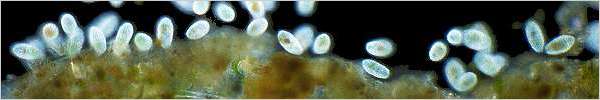
Paramecium.
Paramecium is probably the protozoan most frequently used as an example of a motile single-celled organism in school and university textbooks. Accompanied by the usual
diagram,
it is even sometimes referred to as a "simple" single-celled organism. It can be said here that any single cell which is capable of complex feeding behaviour, digestion and excretion of waste products, reproduction both sexual and asexual, and coordinated movement with chemotaxic and avoidance responses can hardly be regarded as simple -- especially when compared to the limited capabilities of any individual cell of a multicelled animal.
Paramecium was formerly exampled as the classic holotrich ciliate, but in recent reclassifications, it is now variously placed with the Hymenostomata, the Vestibulata or the Nassophorea depending on the actual system.

Movie: 1MB.
Takes about
3 mins to load.
|
A Paramecium foraging in a flocculent mass of organic detritus.
The feeding current created by the cilia of this Paramecium is bringing a continuous stream of food particles to its buccal funnel. It is almost possible to see the vacuoles forming at the extreme end of the funnel. The circulation of the food vacuoles throughout the cytoplasm of the paramecium can be clearly seen.
For some reason, the animation runs very poorly in Internet Explorer 6, but behaves normally (ie. smoothly) in Mozilla.
The fine threads looping about in the background are the filamentous bacterium Beggiatoa.
This video sequence was shot using very simple and cheap equipment.
See the article on videomicrography in the field for details.
Brightfield: x200. |
 |
Paramecium foraging in a gelatinous mass of mucilage-secreting bacteria. A back-and-forth probing action loosens bacteria from the mass, and the currents generated by the cilia draw them into the Paramecium's buccal cavity. The bright granules within the organism are of a sugar-based compound called paramylum (also called paramylon), an energy storage form common to many unicellular organisms.
Darkfield: x200. |
 |
A single Paramecium amongst filamentous bacteria (probably Beggiatoa) and numerous small flagellates, probably Chilomonas. It is somewhat flattened due to pressure from the coverglass caused by evaporation of the specimen.
Darkfield: x300. |
 |
Numerous Paramecia feeding on decaying plant material in a putrefying pond-water sample. The blackened nature of the sample is probably due to sulphides generated by bacteria under low-oxygen conditions. The food vacuoles within the organisms are also black.
Darkfield: x300. |
 |
Numerous Paramecia feeding amongst a tangle of filamentous bacteria and floccules of mucilage-secreting bacteria.
In this and the above picture, the Paramecia are not under pressure from the coverglass, and their characteristic shape can be seen.
Darkfield: x100. |
 |
Same specimen as above at higher magnification. The paired organisms are undergoing conjugation.
Darkfield: x180. |
 |
Same specimen as the two pictures above. A closer view of conjugation -- there is some flattening of the organisms due to coverglass pressure.
This sexual process usually occurs after a long series of asexual mitotic divisions in an ageing population as food reserves become depleted. After exchange of genetic material, the two conjugants will separate and asexual reproduction by mitosis will continue as before (see below).
Darkfield: x300. |
 |
These conjugating Paramecia were found in a sample of pond water which was left to stand, and examined about a week after collection. Very few were noticed in the freshly collected sample, but were present in large numbers in the decaying specimen a week later, along with vast numbers of bacteria, which are their principal food.
The picture shows clearly that the organisms are joined together with their oral grooves aligned. During conjugation (which may take many hours) the macronuclei disintegrate and the micronuclei undergo a complex series of mitotic and meiotic divisions which result in the exchange of DNA between the conjugants. Separation, followed by two mitotic cell divisions of each former conjugant results in eight organisms, each having regained the normal complement of macronucleus and micronucleus.
The paramylum granules of both individuals have collected in a dense mass at the posterior end of the body.
Brightfield: x300. |
 |
Paramecium bursaria is found in similar locations to the Paramecia shown above, but under less polluted conditions (as seen here) is capable of harbouring within its body numerous algal unicells. Taken in by the normal feeding process, they are not digested, but continue to photosynthesize, providing the Paramecium with sugars which supplement its normal diet.
As long as conditions allow, the symbiotic algae replicate within the Paramecium, and when the Paramecium divides, roughly equal numbers of algae will be transferred to each daughter cell.
This commensal or symbiotic relationship with algae is common amongst the ciliates (see also Vorticella and
Ophrydium), and is also found in more complex organisms such as Hydra and marine coral polyps.
Darkfield: x400. |
Coleps.
Coleps is a common holotrich ciliate in freshwater habitats, especially in situations where algae and other plant material has begun to decompose. Their characteristic barrel body shape is due to a semi-rigid pellicle of armoured plates composed of amorphous calcium carbonate. Their bodies are covered with rows of cilia following the longitudinal lines of the plates.
Here is a diagram of Coleps.
 |
A lone Coleps browsing amongst the remains of some deceased microscopic crustacean.
Darkfield, x400. |
 |
Coleps in fairly typical surroundings. The background is of filamentous algae which have begun to decompose and disintegrate. The large food vacuole within the organism's body is green from the algal cell contents which are its food under these conditions.
Darkfield, x400. |
 |
Coleps browsing on decaying plant material. The colour of their food vacuoles gives an indication as to what they have been eating.
Darkfield, x400. |
 |
Coleps feeding among filaments of the alga Oedogonium.
Darkfield, x400. |
 |
This darkfield picture of a somewhat flattened Coleps shows the structure of the calcareous outer pellicle.
A Leitz oil-immersion fluorite objective with integral iris diaphragm was used with a narrow bandpass green filter and fine-grain black and white film to obtain maximum contrast. The substage condenser used was a Beck focusing darkfield condenser. The high magnification is the result of a crop of the original 35mm negative.
A degree of digital contrast enhancement was also employed.
Darkfield: x2000. |
Coleps: Feeding Sequence.
Coleps are scavengers, and the mouth, which is at the forward end of a basket-like pellicle of calcareous plates, can be distended to accomodate quite large meals. They in turn, especially when present in large numbers, form part of the diet of creatures such as worms and predatory protozoa.
Click for some pictures of Coleps at various stages of digestion in the food vacuoles of the ciliate Stentor.
 |
Picture captions anticlockwise from upper left. All pictures are darkfield.
-
A Coleps feeds upon the leaked cell contents of broken filaments of the alga Oedogonium. x200.
-
A single Coleps begins to feed on the body of a recently deceased gastrotrich. The pellicle at the mouth end has expanded to allow the Coleps to engulf its food. x600.
-
The first organism has by now engulfed about a third of the gastrotrich, and is joined by another three individuals who also begin feeding. x600.
-
Feeding continues. The Coleps on the left, which began feeding, has now detatched itself. Others begin to arrive. Three or four Coleps can finish a meal of this size in a period of five minutes or so. x600.
|
 |
 |
 |
|