HOME Editorial Articles Specimen Galleries Tutorials Projects Archive Suppliers Links
Hydrozoa.
Hydra.
Main
Page

1 of 1

Hydra.
Hydra fusca with buds: from Carpenter and Dallinger, 1891. P. 788.


  Hydra.

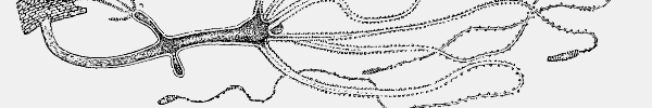
Hydra is included in the phylum Cnidaria, along with sea anemones, jellyfish and coral polyps, and is one of the few freshwater members of this group. One feature all members of the Cnidaria have in common is the possession of thread cells or cnidia or nematocysts -- tiny stinging cells located in the body and tentacles (see diagram) which discharge a paralyzing poison into prey organisms, enabling their capture and ingestion. The nematocysts of Hydra are similar to those of the marine stinging jellyfish which periodically invade beaches, causing injury and sometimes death to humans.

The Hydra's body is a hollow tube consisting of two layers of cells separated by an unstructured gelatinous layer. The outer, clear layer of cells is called the epidermis or ectoderm, and generates the nematocysts. The inner layer is called the endoderm or gastrodermis, and produces the enzymes which digest the Hydra's food. The separating layer is called the mesoglea. It is a gel of various secretions and proteins, containing loose cells not organised into any kind of tissue.
The Hydra's tentacles are hollow, tubular extensions of these three layers.
Here is a diagram of Hydra.

Many members of the Cnidaria establish symbiotic relationships with zoochlorellae which impart a green colour to the polyp. Coral polyps are a well known example of this in the marine environment, and in fresh water, the best known example is Hydra viridis (see below).

Hydra reproduces asexually most of the time by a process of budding, young polyps becoming detatched from the parent when they are fully developed. Seasonal episodes of sexual reproduction also occur, mature polyps developing gonads on the external body wall. Fertilized eggs give rise to tiny planula larvae which swim away, attatch themselves and develop into polyps which continue to reproduce by budding.

 
  Hydra fusca.

Hydra. Hydra fusca: extended polyp with a single bud.
Darkfield: x25.
Hydra. Hydra fusca. This polyp has two buds; the one on the lower left is fairly well developed, but with contracted tentacles, and the one on the right is a new bud, yet to develop tentacles. The small lumps on the tentacles of the parent are the nematocysts, which discharge on contact with a passing prey organism, inducing paralysis and allowing the tentacles to bring the prey to the mouth of the polyp.
Careful examination of the body and two of the tentacles of the adult reveals the tiny protozoan Trichodina ( see diagram ), said to be parasitic upon the Hydra, and usually in a state of continuous gliding movement over the Hydra's body.
Darkfield: x25.
Hydra disgorging water flea. This Hydra has just disgorged the indigestible parts of the body of a water flea, the remains of a previous capture. Hydras have no digestive system or anus as such, and waste products must be ejected through the mouth in a reversal of the process which captured them in the first place.
Darkfield: x25.

 
  Hydra viridis.

Hyra viridis is usually bright green. The colour is due to large numbers of green algal cells, or zoochlorellae, dispersed throughout the entire endoderm, the innermost of the two cell-layers forming the Hydra's tubular body.
The zoochlorellae, as they do in the other creatures which harbour them, use sunlight to produce sugars and generate oxygen, both of which benefit the hydra (however, see below for a comment on commensal algae and coral bleaching).
The metabolic activities of the hydra generate carbon dioxide which is required by the algae. Because they have their own internal garden, green hydras can survive for long periods between prey captures.

All the pictures below were shot with a Kodak DC4800 digital camera.

Hydra viridis on Lemna Hydras are often found hanging from the underside of the floating leaves of various pond plants. Here a Hydra viridis is seen suspended from the underside of a rootlet of the lesser duckweed, Lemna minor.
Incident Light: x25.
Hydra viridis on Lemna. Another shot of the same specimen as the picture above.
Incident Light: x25.
Hydra viridis contracted. Hydra viridis beginning to extend again after going into a fully contracted state.
Incident Light: x100.
Hydra viridis A single polyp of Hydra viridis. Of the two layers of cells which make up the body of a Hydra polyp, the outer clear layer (the ectoderm) is the one which contains the nematocysts, or stinging cells. The inner layer (the endoderm) is the one in which the symbiotic zoochlorellae are found. This double-layered tubular structure is found in both the trunk and tentacles of the polyp, seen here in a semi contracted state.
Brightfield: x100.
H. viridis body. A moderately magnified picture of the partially contracted body of H. viridis showing the distribution of zoochlorellae in the endoderm.
Brightfield: x400.
H. viridis tentacle. A highly magnified picture of the extended tentacle of H. viridis. The nematocysts are produced in the clear outer layer of the ectoderm by specialized cells called cnidoblasts. Also seen are the extended fine hairs (cnidocils) which trigger the nematocysts when they are brushed by passing organisms.
The triggering is believed to be due to a sudden increase in fluid pressure within the nematocyst, causing a fine hollow tube (the thread of the thread cell) to evert, simultaneously injecting a poison into the prey organism. The thread is barbed at its lower extremity, and the holding power of hundreds of discharged nematocysts, together with the effects of the injected toxins, serve to capture surprisingly large and active organisms.
Brightfield: x700.
H. viridis tentacle. Another highly magnified picture of the extended tentacle of H. viridis. In the clear layer of ectodermal cells, the structures of the nematocysts can be seen, as can the cnidocils which trigger them.
Brightfield: x700.

 
A Footnote on Coral Bleaching.

Under favourable conditions, the coral polyps of the world's marine reefs share a very similar relationship to the above with their own commensal algae. At the higher sea temperatures now being experienced by many coral reefs, the algae photosynthesize at an elevated rate, and the higher levels of oxygen produced begin to poison the polyp, which responds by ejecting increasing numbers of its algae. This imbalance results in the eventual rejection of all the algae -- hence the bleaching -- and the death of the polyp.
This is a serious problem indeed, for which there seems no immediate solution.


Click to compose email