HOME Editorial Articles Specimen Galleries Tutorials Projects Archive Suppliers Links
Algae.
Diatoms. 
Page
1 of 2
Algae: Intro. Colonial Diatoms Filamentous Desmids Motile


Quoted from National Geographic: Diatoms, February 1999, P. 117.

  Diatoms.

Diatoms: Page 1 Page 2

Diatom (Arachnoidiscus): from The Microscope; Carpenter and Dallinger, 1891. For much of the course of life on Earth, these microscopic algae have been, and are still, extremely important. More than 10,000 living diatom species are known, with about the same number of named fossil forms.

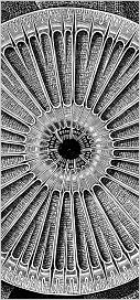
Over ninety percent of the biosphere is plant life, of which diatoms make up about a quarter by weight. They are enormously abundant in the upper layers of the world's oceans, providing high-grade nutrition to creatures as diverse as protozoans and baleen whales, and supplying the atmosphere with around a quarter of its oxygen.
Most diatoms are much less than half a millimetre in size, but their oil-rich, silica-shelled bodies, sinking to the ocean floor in vast numbers over long periods of time, have been transformed into the petroleum deposits of the world, and their skeletons have formed thick strata of diatomaceous earth which has found application in human products as varied as dynamite and (in earlier times) toothpaste.

Whilst most diatoms are to be found in the oceans, they are also abundant and important in freshwater habitats and in moist soil. The siliceous skeleton common to all varieties is frequently described as structured like a pill box or Petri dish, and offers two possible views -- the valve view (as in viewing a Petri dish from the top) and the side or girdle view. They present a great variety of shapes (mostly symmetrical) in valve view, but most commonly are rectangular in girdle view (See Cocconeis below). In general terms, the shape of the valve can be described as pennate (elongated) or centric (circular). The shape and markings of the valve are the means by which species are identified.

The photosynthetic pigment of diatoms is brown, and occurs in the pennate diatoms usually in the form of two identical plastids running the length of the cell (see Navicula below), and in the centric diatoms in the form of numerous sometimes clumped granules.
In the summer waters of a healthy pond, diatoms can grow to such numbers that submerged plants can have the appearance of being covered with a brown mud which the microscope reveals as a dense growth consisting entirely of diatoms.
Notes on diatom reproduction will be added at a later date.

 
  Cocconeis and Gomphonema.

Cocconeis on an algal filament. Two diatoms of the genus Cocconeis are seen in profile (girdle view) clinging to a filament of algae.
Darkfield, x1000.
Cocconeis on an algal filament. A single diatom of the genus Cocconeis seen in profile attatched to a filament of algae.
Darkfield, x1000.
Cocconeis on an algal filament. A Cocconeis diatom attatched to a filament of algae seen from above with the narrow beam of light from the darkfield condenser arranged for maximum visibility.
Darkfield, x1000.
Cocconeis on Lemna rootlet. Two Cocconeis diatoms are seen attatched to a rootlet of the common duckweed (Lemna minor).
Darkfield, x1000.
Gomphonema on a filament of algae. Two diatoms of the genus Gomphonema are seen in girdle view attatched to a filament of the alga Cladophora.
Darkfield, x600.
Many Gomphonema diatoms on a filament of algae. A filament of the alga Cladophora is seen populated with Gomphonema diatoms.
Darkfield, x300.

 
  Navicula.

Navicula diatom on organic matter. A diatom of the common genus Navicula is seen against a background of flocculent organic matter. The two longitudinal brown plastids characteristic of many pennate diatom species are clearly seen. Under the microscope, Navicula (in common with many other diatoms having a raphe) can be seen gliding slowly and erratically in contact with the slide or coverglass. A raphe is a narrow slit running the length of some diatoms through which a slime produced by the diatom is exuded. This partly explains the motion. None of the centric diatoms possess a raphe or show motility.
Darkfield, x600.

This short video sequence (470KB; takes a minute and a half to load) shows a Navicula diatom showing typical movement (made rather more jerky by the low frame rate of the video).
Phase contrast, x1500.
Navicula on organic matter. A dead Navicula diatom imaged in phase contrast, clearly showing the markings on the frustule. The green filament immediately above is (most likely) of the blue-green alga Oscillatoria.
Phase Contrast. x1500.
Navicula diatom amongst debris. Another dead Navicula diatom, this time shown in brightfield illumination.
Click for a diagram of Navicula.
Brightfield, x1500.
Navicula diatom amongst debris. Large numbers of this (living) Navicula diatom was found in a small pond amongst various filamentous green algae along with a variety of browsing ciliates. The bodies of many of the larger ciliates were filled to capacity with these diatoms.
The two plastids are clearly seen.
Phase Contrast, x1800.


Click to compose email