 |
Rotifers:
The Sessile Rotifers. Tube Building in Floscularia. |
Page 2 of 2 |
|
|
Rotifers:
The Sessile Rotifers. Tube Building in Floscularia. |
Page 2 of 2 |

Floscularia feeding amongst filamentous algae.
The following account of tube building by the sessile rotifer Floscularia is taken from The Microscope; by Jabez Hogg, published by George Routledge and Sons, London, 1883 (Pages 460-1). In the preceding text, Hogg has quoted Linnaeus' description of the rotifer's corona as "not unlike a flower of four unequal petals". He goes on to relate Gosse's Carmine is "a beautiful red or crimson pigment obtained from cochineal" (OED). 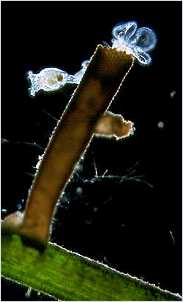 "Below the large petals on the ventral aspect, and just
above the level of the projecting respiratory tubes, is a
small circular disc or aperture, within the margin of which
a rapid rotation goes on. This little organ, which seems
to have hitherto escaped observation, Mr. Gosse can
compare to nothing so well as to one of those little circular
ventilators which we sometimes see in one of the upper
panes of a kitchen-window, running round and round, for
the cure of smoky chimneys.
"Below the large petals on the ventral aspect, and just
above the level of the projecting respiratory tubes, is a
small circular disc or aperture, within the margin of which
a rapid rotation goes on. This little organ, which seems
to have hitherto escaped observation, Mr. Gosse can
compare to nothing so well as to one of those little circular
ventilators which we sometimes see in one of the upper
panes of a kitchen-window, running round and round, for
the cure of smoky chimneys.
The gizzard, or muscular bulb of the gullet, is always very distinct, and its structure is readily demonstrated. It consists of two sub-hemispherical portions, or jaws, each of which is crossed by three developed teeth, which are succeeded by three or four parallel lines, as if new teeth might grow from thence. The teeth are straight, slender, swelling towards their extremity, and pointed. These armed hemispheres work on each other, and on a V-shaped or tabuliform apparatus beneath, common to most of the Rotifers, but in this genus very small. The pellets composing the case are very regular in form and position: in a fine specimen, about the 1-28th of an inch in length when fully expanded, of which the tube was the 1-36th of an inch, Mr. Gosse counted about fifteen longitudinal rows of pellets at one view, which might give about thirty-two or thirty-four rows in all. In November, 1850, Mr. Gosse found a fine specimen attached to a submerged moss from a pond at Hackney; this he saw engaged in building its case, and at the same time discovered the use of the curious little rotatory organ on the neck. When fully expanded, the head is bent back at nearly a right angle to the body, so that the disc is placed nearly perpendicularly, instead of horizontally; the larger petals, which are the frontal ones, being above the smaller pair.  Now, below the large petals (that is, on the
ventral side) there is a projecting angular chin, which is
ciliated; and immediately below this is the little organ in
question. It appears to form a small hemispherical cup,
and is capable of some degree of projection, as if on a short
pedicle.
Now, below the large petals (that is, on the
ventral side) there is a projecting angular chin, which is
ciliated; and immediately below this is the little organ in
question. It appears to form a small hemispherical cup,
and is capable of some degree of projection, as if on a short
pedicle.
On mixing carmine with the water, the course of the ciliary current is readily traced, and forms a fine spectacle. The particles are hurled round the margin of the disc, until they pass off in front through the great sinus, between the larger petals. If the pigment be abundant, the cloudy torrent for the most part rushes off, and prevents our seeing what takes place; but if the atoms be few, we see them swiftly glide along the facial surface, following the irregularities of outline with beautiful precision, dash round the projecting chin like a fleet of boats doubling a bold headland, and lodge themselves, one after another, in the little cup-like receptacle beneath. Mr. Gosse, believing that the pellets of the case might be prepared in the cup-like receptacle, watched the animal, and presently had the satisfaction of seeing it bend its head forward, as anticipated, "and after a second or two raise it again; the little cup having in the meantime lost its contents. It immediately began to fill again; and when it was full, and the contents were consolidated by rotation, aided probably by the admixture of a salivary secretion, it was again bent down to the margin of the case, and emptied of its pellet". This process he saw repeated many times in succession, until a goodly array of dark-red pellets were laid upon the yellowish-brown ones, but very irregularly. After a certain number were deposited in one part, the animal would suddenly turn itself round in its case, and deposit some in another part. It took from two and a half to three and a half minutes to make and deposit a pellet". The darkfield pictures below show details of the corona, antenna and brick-making organ.
|


