HOME Editorial Articles Specimen Galleries Tutorials Projects Archive Suppliers Links
Spiders.
A General Introduction.
Terrestrial Spiders.
Main
Page

1 of 1

Leg of garden spider.
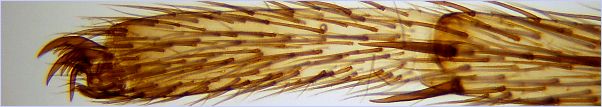
Leg of a garden spider (balsam mounted specimen). Argyopidae aranea.


  Spiders.

The spiders are the best known members of the class Arachnida, which also includes the mites, ticks, scorpions and other scorpion-like creatures.
The bodies of all of the Arachnida have two main parts; the prosoma (or cephalothorax) and the opisthosoma (or abdomen). The prosoma bears six pairs of appendages: the chelicera (jaws), the pedipalps, and four pairs of legs. The chelicera are located forward of the mouth, and are used for holding, piercing, and the injection of poisons which paralyse the prey organism -- usually insects or other spiders.
The spiders are very diverse in size, lifestyle and adaptation. Something like 40,000 species have been described, and there remain many regions on Earth where studies of spiders have not yet been undertaken.

Spiders, like all arthropods, grow in size by a process of moulting, and a mature spider will have moulted between two and twelve times during its lifetime. Females take longer to mature than males, and undergo a larger number of moults.

A spider which is ready to moult usually finds a quiet place to hang by its claws, and by increasing the blood pressure in the prosoma, causes the old cuticle to crack in a line which runs around the prosoma above the level of the legs, and below the eyes.
The spider is then able to slowly extract its legs from the cuticle by alternately increasing and releasing its blood pressure. The thin cuticle covering the abdomen also splits and contracts, leaving only a shrivelled remnant.
The newly-emerged spider, soft and vulnerable, hangs by a thread intil its new cuticle hardens.

The series of pictures below shows the discarded cuticle, or exuviae, of a common house spider.

1. Spider cuticle. The discarded cuticle of a common spider. It would be wrong to say that this is a perfectly detailed replica of the external features of the spider which originally inhabited it, because it's not a replica -- it's the real thing.
Brightfield, x8.
2. Spider cuticle. A closer view shows that the dorsal half of the prosoma has flipped over like a lid on a hinge, revealing its inside surface. Between the two parts of the prosoma is the shrivelled remnant of the cuticle which once covered the opisthosoma (abdomen).
Brightfield, x16.
3. Spider cuticle. Here, in dorsal aspect, the details of the chelicera and pedipalps are clearly seen, as are the hollows from which the spider has extracted its legs. The stalk seen extending from between the celicera is the dried remains of the lining of the oesophagus and upper digestive system, also shed during a moult.
Brightfield, x32.
4. Spider cuticle. Same field as the picture above, but viewed in ventral aspect, giving more detail of the mouth parts, pedipalps, sternum and upper leg segments.
Brightfield, x32.
5. Spider cuticle. The cuticle of the prosoma (cephalothorax) showing the eye lenses in profile. On an analogy with the mammalian eye, the thin domed structures seen here could be described as the cornea of the spider's eye. During the moult, the vitreous body which forms the main part of the lens remains with the spider.
Here is a diagram showing the structure of a spider's eye.
Brightfield, x50.
6. Spider cuticle. A closer view of the cuticle of the eye lenses. The dent in the central eye is a visual indicator of the thinness of the cuticle.
Brightfield, x70.

The above photographs were taken using a Kodak DC4800 3.1 Megapixel digital camera fitted to the eyepiece of a low-power Russian MBC2 stereo microscope.

 
  A Newly Hatched Spiderling.

These pictures of a young spider, recently hatched from its egg, were taken using a microscope illuminated by a double electronic flash setup which is described as part of the article on electronic flash as a microscope illuminant. Click here for more details on the technique used.

The pictures show the spider suspended in a web constructed across the lower half of a small transparent plastic container, and provide an opportunity to observe the way in which the creature uses its rear legs to manipulate the thread generated by the spinnerets.


1. Young spider.


2. Young spider.
Picture captions anticlockwise from upper left. All pictures are Rheinberg with incident light fill-in.
  1. The spider seen in dorsal view, suspended in its web. x40.
  2. Another dorsal view of the spider in its web. x40.
  3. Here the spider is seen in ventral aspect, hanging upside down in its web. The claws on each of its legs provide a secure hold on the threads of the web, which are not sticky. x40.
  4. A view from behind shows the rear of the abdomen, and a thread of silk coming from the spinnerets. x60.
  5. This picture shows the way in which the two rear pairs of legs are involved in the manipulation of the thread from the spinnerets. The last pair of legs in particular are seen holding the newly-formed threads prior to attatching them to other threads of the web. x60.
  6. The web itself. Many spiders weave apparently haphazard nets of this general apppearance, but perhaps this spider would have done a neater job had it not been blitzed with an electronic flash a dozen times in quick succession.
3. Young spider. 4. Young spider. 5. Young spider. 6. Young spider's web.